Iron Deposits in Lung Macrophages May Be Sign of Microvascular Problems in IPF Patients


Increased deposits of iron in immune cells called macrophages were found to be independently correlated with pulmonary vascular resistance (PVR) in patients with idiopathic pulmonary fibrosis (IPF), according to a recent study.
Findings of hemosiderin-laden (an iron-storage complex within cells) in macrophages in the lungs of IPF patients might also be an early sign of events linked to microvascular abnormality, it reported.
The study, “Hemosiderin-laden macrophages are an independent factor correlated with pulmonary vascular resistance in idiopathic pulmonary fibrosis: a case control study,” appeared in the journal BMC Pulmonary Medicine.
Researchers have commonly used findings of hemosiderin deposits or exaggerated numbers of hemosiderin-laden macrophages (HLM) in bronchoalveolar lavage fluid (BALF) as a diagnostic biomarker of alveolar hemorrhage. Recently, this macrophage alteration has also been reported in IPF cases. Hemosiderin deposits in lung tissue is linked to pulmonary hypertension.
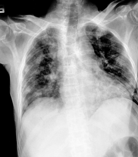
The study is a retrospective review of initial evaluation information on 103 patients with mild to moderate IPF, who underwent lung biopsies at Japan’s Tosei General Hospital between November 2007 and March 2014. Most of these IPF patients were males and smokers, with a median age of 66.
Researchers were interested in determining whether the number of hemosiderin-laden macrophages in the patients’ bronchoalveolar lavage fluid, as determined by an HLM score, correlated with PVR, or vascular resistance, in IPF. This score consists of dividing the number of macrophages with iron deposits by the total number of macrophages.
Results found that the HLM score was positively correlated with two pulmonary hemodynamic parameters, the mean pulmonary arterial pressure (mPAP) and PVR. They then evaluated mPAP and PVR by right heart catheterization protocol, and observed the same correlation between PVR with HLM scores, confirming previous observations.
“This is the first report to show a significant correlation between pulmonary hemodynamic parameters measured by RHC [right heart catheterization] and HLM in BALF in patients with IPF,” the authors wrote.
The specific BALF cellular pattern of IPF is not fully understood. However, this evaluation can be performed as a means to identify IPF — or exclude other conditions — for differential diagnoses. Evaluating HLM scores, which might correlate with PVR, can also provide more on the BALF status of IPF patients. This may help detect pulmonary hypertension in these patients in the disease’s early phase.
“Among the data from patients with idiopathic pulmonary fibrosis, hemosiderin-laden macrophages in bronchoalveolar-lavage fluid were significantly correlated with pulmonary hemodynamic parameters evaluated by right heart catheterization, and were an independent correlating factor of pulmonary vascular resistance,” the team concluded, adding that “hemosiderin deposition in the lungs of IPF is already significant even in mild to moderate IPF, and HLM score can reflect such early microvascular abnormality.”


