Umbilical Cord Stem Cells Help Limit Inflammation in Mice

unoL/Shutterstock
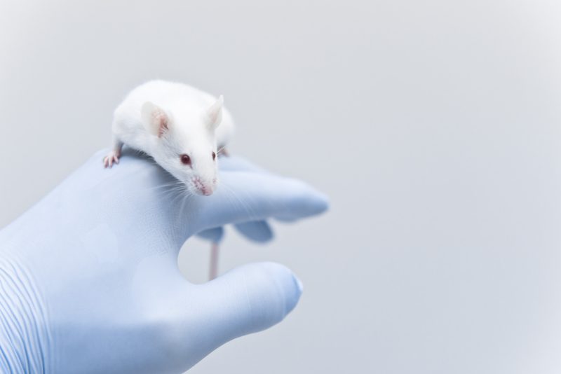
Stem cells found in human umbilical cords limited lung damage associated with pulmonary fibrosis (PF) by reducing an excessive inflammatory immune response, a recent study of mice has found.
Understanding how these stem cells interact with the immune system could improve future PF cell therapies, the researchers noted.
The study, “Human umbilical cord mesenchymal stromal cells attenuate pulmonary fibrosis via regulatory T cell through interaction with macrophage,” was published in the journal Stem Cell Research & Therapy.
The lung scarring, or fibrosis, that characterizes PF results from the disproportionate buildup of extracellular matrix components like collagen. Over time, this collagen-dense scar tissue causes lungs to become stiffer and lowers their ability to exchange gas. (The extracellular matrix is the network of proteins and other molecules that surrounds and supports cells in tissues.)
Recently, mesenchymal stromal cells (MSCs) have drawn scientists’ interest for their potential to decrease collagen deposition and therefore fibrosis. These cells have demonstrated an ability to limit inflammatory immune reactions in PF animal models. Yet, exactly how they accomplish this is still not clear.
In an effort to learn more, a group of scientists from Shenzhen, China, investigated how MSCs-derived human umbilical cords interact with the immune system in a mouse model of induced PF.
After treating animals with the anti-cancer medication bleomycin to trigger the onset of PF, researchers administered human umbilical cord-derived MSCs (hucMSCs) to one group of mice and compared their responses with those of untreated animals.
Treated mice lived longer than their untreated peers, showing fewer signs of collagen buildup in the lungs. Furthermore, fibroblasts — connective cells associated with fibrosis — proliferated less in treated mice, while AT2 cells, which play a key role in lung tissue repair, increased more.
These effects combined suggested that treatment with hucMSCs could limit the extent of fibrosis in PF, as has been seen in past studies.
Investigators then explored how MSCs might accomplish their therapeutic effects, often referred to in scientific literature as their mechanism of action.
They found that macrophages — specialized immune cells that trigger inflammation upon detecting and destroying harmful organisms — would engulf, or “swallow,” hucMSCs. This caused them to increase the expression, or activity, of a specific set of genes.
One of these genes, Cxc10, led to the recruitment of regulatory T-cells (Tregs) to the lungs. Tregs are known to suppress the body’s immune response, preventing it from spiraling out of control and causing unintended tissue damage.
“Our study establishes a link between hucMSCs, macrophage, Treg, and PF,” the researchers wrote. “It provides new insights into how hucMSCs interact with macrophage during the repair process of bleomycin-induced PF and play its immunoregulation function.”


