
Calcium Signals Behind Conversion of Healing Cells into Promoters of Fibrosis, Study Finds
Written by |
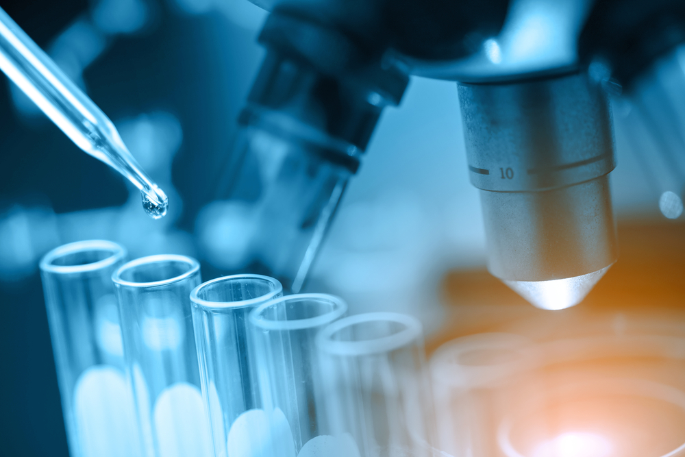
New research provides important insights into the process of activation and formation of cells that are crucial for fibrosis to occur, uncovering potential targets that can help control fibrosis in chronic diseases such as pulmonary fibrosis (PF).
Scientists at the Lewis Katz School of Medicine at Temple University (LKSOM) discovered that certain metabolic pathways involved in calcium signals play a central role in rewiring cells to convert into myofibroblasts, essential players in fibrosis.
The discovery was done in animal and cellular models, so it is still at an early stage. However, its researchers hope the new pathways identified can be manipulated to one day treat fibrotic diseases.
Their findings are described in the study “Mitochondrial calcium exchange links metabolism with the epigenome to control cellular differentiation,” published in the journal Nature Communications.
Fibroblasts are specialized cells that are attracted to sites of injury, where they proliferate and help to repair damaged tissues. In the process, fibroblasts give rise to healing cells called myofibroblasts. In normal situations, these cells are essential in the wound healing and regeneration process, after which they are naturally removed from the affected areas.
But myofibroblasts that are excessively activated, and stay in tissues longer than would be normal, can give rise to pathological situations marked by excessive scarring, as happens in fibrotic diseases like PF, liver cirrhosis, and heart failure.
Given their important role in health and disease, LKSOM researchers were interested in understanding more deeply the mechanisms underlying myofibroblast formation, a process still unclear to scientists in many aspects.
Carrying out experiments in mouse models and in fibroblasts growing in the lab, researchers found that stress-induced changes in calcium uptake by the mitochondria (the cell’s powerhouses) drives the conversion of fibroblasts into myofibroblasts.
When mitochondrial calcium levels drop, a series of coordinated changes in the metabolism of fibroblasts begin that result in the increase of a molecule called alpha-ketoglutarate. In turn, this leads to a genetic reprogramming of fibroblasts, changing the genes that are highly or lowly turned “on” in a way that triggers the differentiation of myofibroblasts.
“Our experiments show that the accumulation of fibrotic cells results directly from mitochondrial calcium-dependent genomic reprogramming involving α-ketoglutarate, which changes the structure of chromatin, or DNA packaging — a phenomenon referred to as epigenetics and that is important in regulating gene expression,” John W. Elrod, PhD, a professor at LKSOM and the study’s senior investigator, said in a press release.
“The key finding, that a change in mitochondrial calcium uptake plays a central role in metabolic and genetic reprogramming, presents new opportunities for investigation,” Elrod added. “We hope that the new pathways we’ve identified as essential to myofibroblast formation can be manipulated to treat fibrotic disease.”
The team plans to investigate further the role of metabolism in fibrosis, and to conduct more genome-wide sequencing studies to better understand the changes in DNA structure that underpin the prolonged presence of myofibroblasts in tissues during disease.


Leave a comment
Fill in the required fields to post. Your email address will not be published.