Cells Surrounding Small Blood Vessels in the Lungs May Play a Role in IPF Progression, Study Finds


Cells present in the outer rims of small blood vessels in the lung, called pericytes, convert from a healthy state into a fibrotic one, promoting the progression of pulmonary fibrosis, new findings suggest.
The discovery shows how pericytes respond to the microenvironment in the lungs, especially the mechanical changes in their surroundings, to promote disease. Moreover, the therapeutic effects of treatments like Ofev (nintedanib), marketed by Boehringer Ingelheim, may include a previously unknown action on pericytes.
The study, “Human pericytes adopt myofibroblast properties in the microenvironment of the IPF lung,” was published in the Journal of Clinical Investigation Insight.
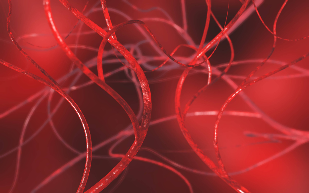
A research team led by Angelica Gonzalez at Yale University is focusing on small blood vessels, or capillaries, that are present in organs including the lungs, brain, and heart and whose surface is wrapped by pericytes.
“We’re looking at these vessels because when IPF [idiopathic pulmonary fibrosis] gets pretty bad, those small vessels go away and the tissue dies because there is limited nutrient and oxygen delivery to it,” Gonzalez, Donna L. Dubinsky Associate Professor of Biomedical Engineering at Yale, said in a press release.
Research into the function of pericytes in PF has lagged, and scientists have long debated what exactly happens to these cells.
Looking at lung samples from IPF patients, Gonzalez and her team found that pericytes made up 15 to 21 percent of the fibrotic lesions seen.
“These findings were the first identification anyone’s made that pericytes make up a portion of these lesions,” Gonzalez said. “No one before had confirmed that these cells could be contributing to the disease. Now we see that these blood vessel cells are still alive – they just become dysfunctional, as opposed to being supportive of healthy tissue.”
The team then developed an engineered human lung and infused pericytes into IPF or control lungs and cultured each for seven days. They observed that pericytes grown on IPF lungs adopted the features of the α-smooth muscle actin expressing myofibroblasts, the cells responsible for the synthesis and buildup of scar tissue (fibrosis).
They performed further experiments and found that pericytes changed their behavior in response to the lung microenvironment, particularly to the mechanical changes of the surrounding tissue. This process, called mechanosensing, is the main trigger for the transformation of pericytes from healthy cells to fibrosis-promoting cells, researchers found.
“These data indicate that even a small increase in substrate stiffness is sufficient to promote myofibroblast-like characteristics in human PC [pericytes] via mechanotransductive signalling,” researchers wrote.
Next, they set out to discover whether common IPF therapeutics, such as Ofev, had any effect on the fibrosis-promoting pericytes. Cells were first treated with TGF-beta1 — the central mediator of fibrosis that regulates the fibroblast-to-myofibroblast transition — followed by treatment with Ofev for an additional seven days. Results showed that Ofev treatment reduced the levels of collagen fibronectin, key components of scarring tissue and stiffness in the fibrotic lung.
“We hit the cells with this drug, and they started producing enzymes that degrade the environment and make it soft again,” Gonzalez said. “These data demonstrate a potentially novel and previously unrecognized mechanism of action of nintedanib that involves the regulation of ECM [extracellularmatrix] production by PC [pericytes]-derived myofibroblasts.”
These findings suggest that Ofev, previously thought to exert its therapeutic effects by acting on resident lung fibroblasts, may also affect pericytes.
As tissue scarring is not only restricted to the lungs, Gonzalez will now assess the role of pericytes in other organs, including the skin and the heart.


