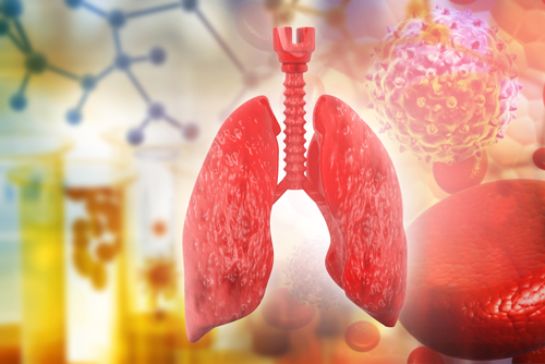
Low Levels of HOPX Protein in Lung Impair Cell Regeneration, Worsen IPF, Study Suggests


Low levels of the homeobox only protein x (HOPX) in lung samples from idiopathic pulmonary fibrosis (IPF) patients are linked to lung function decline and disease progression, a new study suggests.
The study, “Dynamic expression of HOPX in alveolar epithelial cells reflects injury and repair during the progression of pulmonary fibrosis,” was published in the journal Nature Scientific Reports.
During development of the lungs, HOPX is important for the formation of the distal lung — the part of the respiratory system that includes tiny “tree branch” airways called bronchioles and the densely packed honeycomb alveolar air sacs that regulate gas exchange.
In lungs of adult mice, HOPX is found in a group of epithelial cells in the alveoli. Previous research has shown that HOPX-positive cells help regenerate the alveoli after a trauma, such as surgical removal of all or part of the lung (pneumonectomy).
However, the function of HOPX-positive cells in adult fibrotic lung diseases, including IPF, is unknown.
Researchers evaluated HOPX expression during in vitro transition of alveolar epithelial type 1 cells into type 2 — the two main cell types that constitute alveoli epithelium. The type 1 cells are the gas exchange surface cells in the alveoli. The type 2 cells act as “caretakers” of the alveoli, producing protective molecules and replenishing the alveoli cell content when needed.
The Pulmonary Fibrosis News forums are a place to connect with other patients, share tips and talk about the latest research. Check them out today!
They found that HOPX levels were increased during the in vitro transition between the two cell types, and this increase was also detected in vivo in the alveolar epithelium of an IPF mouse model.
Decreasing the levels of HOPX in a mouse’s alveolar epithelial cell line suppressed the transition of alveolar epithelial type 1 into type 2 cells, suggesting that HOPX is involved in the suppression of alveolar epithelial cells’ proliferation.
Researchers then analyzed lung samples of IPF patients and measured the levels of HOPX compared to healthy controls. In contrast to the results in mice, HOPX expression was significantly lower in the IPF lungs compared with control lungs. Moreover, the decrease had a significant correlation with a decline in lung function and progression of IPF.
The findings suggest that HPOX levels are potentially increased during early alveolar injury and repair process in the lung but its lower expression in later stages of IPF may be to blame for failed regeneration, promoting disease progression.
“HOPX contributes to alveolar injury/repair process in fibrotic lung diseases, but fails to regenerate alveolar epithelium in IPF, due to loss of HOPX. HOPX may thus be a potential indicator of the progression of fibrosis,” the researchers said.


