Lung CT Scans May Help Predict Mortality Risk of IPF Patients


Lung evaluations via a CT scan might help predict the mortality risk in idiopathic pulmonary fibrosis (IPF) patients more accurately than the decline in forced vital capacity, a study shows.
The study, “Serial CT analysis in idiopathic pulmonary fibrosis: comparison of visual features that determine patient outcome,” was published in the journal Thorax.
Anti-fibrotic treatments, aimed at preventing lung scarring, can help slow the lung function decline of IPF patients. Forced vital capacity (FVC), a test that evaluates the amount of air forcefully exhaled after taking a deep breath, is commonly used to monitor lung function in IPF.
Since the introduction of anti-fibrotic medications, fewer IPF patients experience a decline in FVC of 10% or more per year; in fact, most experience between 5% and 9.9% annualized FVC declines.
Small variations in FVC make it harder to monitor disease, and there can be some question as to whether changes in these measurements actually correspond to disease worsening.
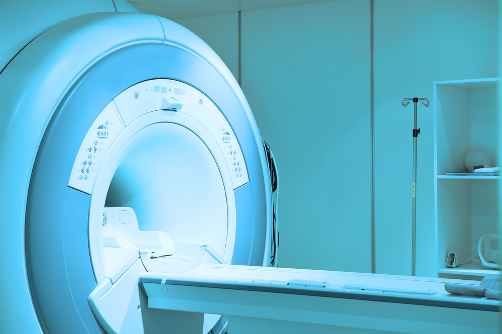
Lung CT, considered a complementary tool to FVC, can be used to determine whether changes in FVC reflect actual lung function decline.
Researchers at University College London and their colleagues investigated how changes in CT visual parameters correlate with IPF disease progression using two groups of patients: the first included 103 patients with IPF followed at the Royal Brompton Hospital in London, and the second had 108 patients from St. Antonius Hospital in Utrecht, Netherlands; Ege Hospital Izmir in Turkey; and Southampton General Hospital in the U.K.
Age, gender, and lung function at enrollment (baseline) were similar between the two groups. CT visual parameters were evaluated by radiologists and scored using a five-point scale.
Considering each parameter individually, the researchers found that several factors were strong predictors of mortality: namely honeycombing extent (patches of cystic spaces), severity of traction bronchiectasis (irreversible dilation of bronchi), and interstitial lung disease extent in the right middle lobe and right and left lower lung lobes.
They then assessed the predictive value of these parameters by adjusting the analysis for patient age, gender, anti-fibrotic use, and baseline diffusing capacity of the lungs for carbon monoxide (DLCO; the ability of the lungs to transfer oxygen from the air to the blood). They observed that traction bronchiectasis severity was the strongest independent factor for mortality prediction.
Compared with FVC decline, this CT-based parameter was a stronger mortality predictor.
In addition, when the researchers divided both groups according to three FVC decline categories — 10% or more (107 patients), 5%–9.9% (53 patients), and minus 5%–4.9% (47 patients) — they saw that traction bronchiectasis severity was able to predict mortality regardless of the FVC decline group.
In particular, for patients with an FVC decline of 5%–9.9%, traction bronchiectasis was a stronger mortality predictor than continuous FVC decline.
These findings showed that the CT parameter traction bronchiectasis severity is a useful tool to monitor IPF disease progression and can more accurately predict mortality.
“Our study has shown that change in traction bronchiectasis is a reproducible measure of disease progression in IPF,” the researchers wrote, adding that it “identifies more patients with disease worsening than honeycombing change.”
According to the team, changes in traction bronchiectasis “can be used to resolve indeterminate FVC declines, which are likely to be seen with increasing frequency in patients with IPF receiving antifibrotics.”


