PDGFA Found to Stimulate Lung Tissue Repair in Aged IPF Mice

Pretreatment with retinoic acid was found to stimulate the repair of damaged alveoli — the tiny air sacs in the lungs — in a mouse model of idiopathic pulmonary fibrosis (IPF) by upregulating a molecule called PDGFA.
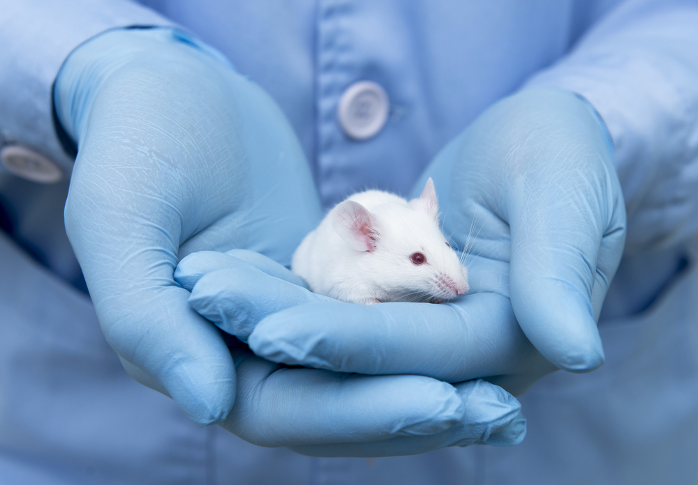
The researchers used an aged mouse model to simulate the lung injuries found in older IPF patients.
Their findings support further development of PDGFA-focused therapeutic strategies that may impact IPF.
The study, “Pretreatment of aged mice with retinoic acid supports alveolar regeneration via upregulation of reciprocal PDGFA signalling,” was published in the journal Thorax.
IPF is characterized by excessive wound healing that leads to scarring and increased stiffness of the lungs. While the mechanisms that underlie this process are unclear, it is thought to occur due to repeated injury to the cells, called epithelial cells, that line the tiny air sacs in the lung (alveoli). It’s also thought to be due to the overactivation of cells known as fibroblasts, which normally generate collagen and extracellular matrix (ECM) components that are essential for normal healing and tissue support.
The body’s failure to regenerate healthy alveoli after injury leads to the loss of alveolar type 1 (AT1) epithelial cells, which are cells that line alveoli where gas exchange occurs between the lungs and the bloodstream. Moreover, mouse models have shown alveoli regeneration failure increases with age.
Lung repair models using mice and rats have shown that treatment with a compound called retinoic acid can restore alveolar regeneration. However, the exact signaling mechanisms of this repair process remain unknown.
To fill this knowledge gap, a team led by researchers at the Cincinnati Children’s Hospital Medical Center, in Ohio, sought to uncover molecules involved in regulating lung and alveolar repair. Along with cell analysis, the team used a lung injury mouse model called PNX, in which part of the lung was surgically removed.
Furthermore, the team grew in vitro (in-the-lab) 3D cell cultures called organoids from epithelial cells isolated from mice or IPF patients. These were cultured along with young (12–16 weeks of age), aged (greater than 40 weeks of age), or retinoic acid-pretreated mouse fibroblasts to validate potentially important repair molecules.
First, aged mice were treated with retinoic acid before PNX surgery and lung tissue was examined after surgery.
“We hypothesised that the treatment of aged mice with [retinoic acid] prior to PNX injury … would promote alveolar regeneration in aged lungs,” the researchers wrote.
The results showed that retinoic acid pretreatment indeed restored alveoli in aged mice. Further experiments revealed the growth of a type of matrix-producing fibroblast with a receptor called PDGFRA played an essential role in alveolar repair.
Changes in gene activity in PDGFRA-fibroblasts associated with alveolar regeneration were assessed after PNX surgery. Both PDGFRA and its binding protein PDGFA were significantly upregulated in fibroblasts and epithelial cells in the retinoic acid-pretreated mice’s lungs compared with aged lungs.
Specifically, in aged PDGFRA-fibroblasts, a large group of genes was altered by PNX injury, while retinoic acid-pretreatment prevented these changes. In contrast, retinoic acid pretreatment of epithelial cells induced genes that were not changed by PNX.
“These data suggest that PNX in aged lungs induces ‘misdirected’ gene activation and inactivation of PDGFRA+ fibroblasts that result in failure to regenerate,” the team wrote. Retinoic acid “prevents these ‘misdirected’ gene changes, allowing regeneration,” they wrote.
Next, experiments to identify gene networks associated with alveolar regeneration in PDGFRA-fibroblasts found a significant increase — by 5.9 times — in PDGFA expression in aged retinoic acid-pretreated PNX mice compared with aged PNX. These results suggest that “increased reciprocal PDGFA signalling is essential for alveolar regeneration after PNX,” the researchers wrote.
To test the role of PDGFA signaling, lung organoids were generated by combining lung epithelial cells with PDGFRA-fibroblasts isolated from young, aged, and retinoic acid-pretreated mouse lungs.
Markers for AT1 alveolar cell production, as well as supportive AT2 cells, were increased in organoids with young or retinoic acid-pretreated lung PDGFRA-fibroblasts, compared with organoids cultured from aged mice.
Additionally, when organoid cultures from aged PDGFRA-fibroblasts were treated with PDGFA ligand, proper growth of epithelial AT1 and AT2 was restored by showing increased cell markers.
Treating organoids generated from aged mouse fibroblasts and human IPF epithelial cells with PDGFA enhanced the proper growth of AT1 and AT2 cells to similar levels found in organoids generated with RA-pretreated or young fibroblasts, suggesting that “epithelial cells from patients with IPF have not entirely lost the ability to differentiate into AT1 cells.”
Finally, to test the importance of PDGFA signaling, organoids were grown from aged mouse epithelial cells co-cultured with young, aged, or retinoic acid-pretreated PDGFRA-fibroblasts.
These organoids were then exposed to a PDGFA-neutralizing antibody, which reduced AT1 and AT2 markers in organoids created with young and aged retinoic acid-pretreated PDGFRA-fibroblasts. In contrast, organoids developed with aged PDGFRA-fibroblasts had low expression of AT1 markers, which demonstrated that “specific inhibition of PDGFA ligand is sufficient to reduce AT1 differentiation,” the researchers wrote.
“Our data support the concept that [retinoic acid] indirectly induces reciprocal PDGFA signalling, which activates regenerative fibroblasts that support alveolar epithelial cell differentiation and repair, providing a potential therapeutic strategy to influence the pathogenesis of IPF,” the team concluded.


