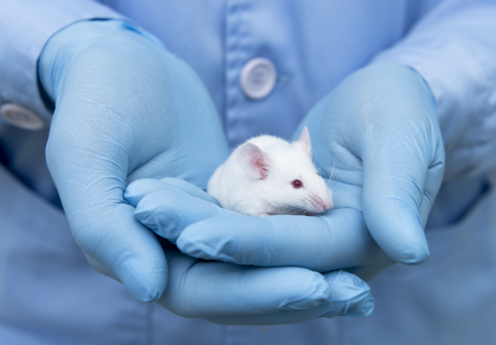
Target FPR-1 Protein or Neutrophils in Fibrotic Lungs, Mouse Study Suggests

The FPR-1 protein is necessary for the recruitment of immune cells, namely neutrophils and inflammatory macrophages, to the lungs where they promote pulmonary fibrosis (PF), a mouse study shows.
Depletion of FPR-1 or neutrophils prevented lung fibrosis and inflammation, supporting their potential as therapeutic targets in PF.
The study “FPR-1 is an important regulator of neutrophil recruitment and a tissue-specific driver of pulmonary fibrosis” was published in the journal JCI Insight.
Neutrophils are key inflammatory cells that play a role in the early stages of wound repair and fibrosis. FPR-1 (formyl peptide receptor 1) is highly abundant in neutrophils, and has been shown to regulate their function. However, whether FPR-1 plays a role in fibrosis remains unknown.
Researchers in the U.K. and Canada investigated the role of FPR-1 in the early stages of fibrosis in multiple organs, including the lung, liver, and kidney. They used wild-type mice as controls, and compared the results to mice genetically engineered to lack FPR-1 (called FPR-1 “knock-out” or KO mice).
Results showed that while control mice developed inflammation and pulmonary fibrosis after being exposed to bleomycin — a commonly used compound to induce PF — the FPR-1 KO mice were protected. Specifically, these animals showed significant lower levels of: collagen deposition, which is produced in excess in fibrotic tissue; activated myofibroblasts, the cells responsible for the synthesis and build-up of scar tissue (fibrosis); and markers of inflammation.
Moreover, 24 hours after bleomycin treatment, lung analysis showed that FPR-1 KO mice had a significantly lower number of neutrophils, detected by the presence of a neutrophil marker called NIMP, compared to control mice — an average of 4.686 cells per field of view versus 9.725 cells in control mice.
A decrease also was seen for inflammatory macrophages (marked by the protein CD68) on day one (11.17 vs. 15.52 cells per field in control mice), and five days following bleomycin (9.157 vs. 17.42 in control mice).
Exposure to the pro-inflammatory molecule TGF-beta 1, however, led to the same degree of fibrosis in both groups of mice.
These results suggest that “FPR-1 is not directly involved in the fibrogenic response in the lung, but instead contributes to inflammation by driving immune cell recruitment and activation in response to tissue damage,” researchers wrote.
Apart from the results in the lung, FPR-1, however, seems to have no role in the early stages or progression of fibrosis in the liver, as researchers saw no differences in control and FPR-1 KO mice after inducing liver damage (with carbon tetrachloride, CCl4 injection) and in two additional models of liver fibrosis.
The same was seen in a model of kidney (renal) fibrosis.
In agreement with these results, researchers saw no differences in the recruitment of neutrophils in the liver or kidney between control and KO mice.
“Mechanistically, we observed a failure to effectively recruit neutrophils to the lungs of [FPR-1 KO] mice, whereas neutrophil recruitment was unaffected in the liver and kidney,” researchers wrote.
Further experiments showed that the presence of FPR-1 on neutrophils is necessary for their lungs to heal following lung injury.
Depleting neutrophils before bleomycin treatment reduced both lung inflammation and fibrosis. This reduction also was accompanied by a lower number of myofibroblasts.
“These data suggest that neutrophils are required for efficient recruitment of inflammatory macrophages to the damaged lung and for the development of fibrosis,” researchers wrote.
Overall, the results support “the potential therapeutic benefit of targeting FPR-1 or neutrophils in the fibrotic lung,” the team concluded.


