
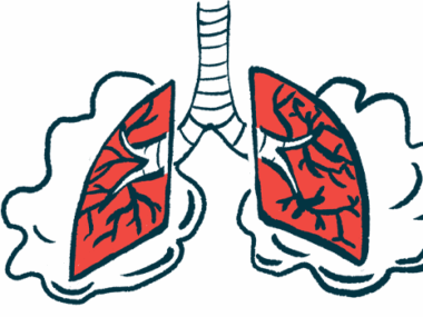
Study finds how extracellular histones drive lung fibrosis
Discovery could lead to new avenues in treatment of pulmonary fibrosis
Written by |

Proteins called histones can trigger lung fibrosis (scarring) by prompting platelets, cell fragments involved in blood clotting, to release a signaling molecule called transforming growth factor beta 1 (TGFB1), a new study found.
TGFB1, in turn, blocks the production of interleukin-27 (IL-27), a signaling molecule that normally helps to limit scarring. Targeting this molecular signaling circuit may open new avenues for treating fibrotic diseases including pulmonary fibrosis.
“Although histones have previously been implicated in fibrosis, how they mediate the development of the disease is not completely understood. Our findings provide novel mechanistic insights into how the altered interactions between different cell types contributes to histone-mediated fibrosis development,” study authors Arjun Sharma and Markus Bosmann, MD, of the University Medical Center of the Johannes Gutenberg-University Mainz, in Germany, said in a university news story.
The study, “Externalized histones fuel pulmonary fibrosis via a platelet-macrophage circuit of TGFβ1 and IL-27,” was published in PNAS.

Histones and DNA
Histones are structural proteins that hold DNA in place. Long strands of DNA are wrapped around histone proteins, similar to thread wrapped around a spool.
Most of the time, histones and DNA are kept securely inside the cell’s nucleus. But as part of certain inflammatory reactions, immune cells called neutrophils sometimes release large tangles of histones and DNA into the extracellular (outside-the-cell) space, which are called neutrophil extracellular traps or NETs. Like a spider web catching flies, NETs can catch infectious invaders like bacteria.
While NETs can be helpful for fighting infections, a growing body of research has suggested that NETs, and in particular extracellular histones, may also work to trigger fibrosis. In fibrotic diseases like pulmonary fibrosis, abnormal activity of immune cells is a major driver of scarring, as immune inflammation and scarring are closely linked biological processes.
In this study, researchers in Germany first analyzed lung fluid from 29 people with idiopathic pulmonary fibrosis (IPF) and 10 people without lung disease. Results showed levels of extracellular histones were significantly higher in the IPF patients.
Pulmonary fibrosis in a mouse model
The team then conducted a series of experiments in a mouse model where pulmonary fibrosis was induced using the chemical bleomycin. They showed that extracellular histones were mainly released by neutrophils.
The researchers further showed that extracellular histones prompted platelets to secrete the signaling molecule TGFB1. This molecule in turn blocked the production of IL-27 by macrophages, another type of immune cell.
Mice that were engineered to lack IL-27 had notably more severe fibrosis, demonstrating that this molecule normally helps to limit fibrosis.
Thus, the work here outlines a molecular mechanism for how extracellular histones can promote fibrosis, by triggering production of TGFB1 and thereby reducing levels of IL-27.
“Externalized histones activate platelets to release TGFβ1, which subsequently antagonizes antifibrotic Interleukin-27 (IL-27) production from macrophages via multiple intracellular signaling pathways,” the scientists concluded.
The researchers further showed treatment with antibodies that block extracellular histones reduced fibrosis in the mouse model, implying this circuit may be a useful target for treatments.
“This study helps bridge a crucial knowledge gap by elucidating the role of three key proteins – histones, TGFβ1, and IL-27 in fibrosis development, opening up avenues for new therapies,” Bosmann said.


Leave a comment
Fill in the required fields to post. Your email address will not be published.